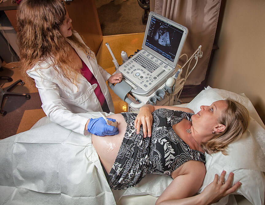
Accredited ultrasound services require a highly educated and skilled expert to operate the ultrasound machine and perform diagnostic examinations. Ultrasound is used both for pregnancy and also for the diagnosis of GYN issues. A physician interprets the resulting images. Based on one or multiple factors, he or she affirms that the anatomy is normal, or if an abnormality is suspected, recommends further testing or treatment.
How Ultrasound Changed Pregnancy
When the technology of ultrasound first emerged, pregnant women were excited to see their developing babies. A patient would lie on a cold table, bladder filled with the large quantity of water she was required to drink to yield a clear picture. The technician lubricated her stomach with cold gel, and proceeded to rub a transducer probe across her belly while the anxious future mom and dad looked on.
The pictures were often disappointing – blurred and grainy. Without the sonographer patiently tracing the image with a finger on the monitor, prospective pareents didn’t know if they were admiring their baby’s face or the uterus it was inside. As for gender, well, that was still a hit and miss thing – basically no different from the baby shower tradition of swinging a ring on a rope, and guessing the baby’s sex based on whether the ring twirled or swung like a pendulum.
Back then, only the sonographer’s experienced eye could differentiate the important information from the blips and blotches on the monitor to insure that all was well. But since then, the quality of ultrasound – and the training of technicians – have evolved a hundredfold.
Briefly, How Does Ultrasound Work?
The ultrasonographer sends harmless high frequency sounds, undetected by human ears, toward a ‘target,’ in this case, your growing baby. Those sound waves are directed via a probe called a transducer. The sound waves bounce back, creating an ‘echo’ when they encounter the bone or tissue of the baby. That echo creates an image on a monitor—a picture of your baby complete with all its parts, including internal bones, tissue, organs and even blood flow. You may recognize only larger parts of your baby, like face, limbs, heart, etc., but the technician knows what all the little pieces of the fetus are. He or she is trained to decipher and measure every part to verify due date, gender, and to detect if anything is out of the ordinary.
Chief Ultrasonographer Discusses Ultrasounds
Cherokee Women’s Health’s chief ultrasonographer Brenda Peters talks about her experience performing ultrasounds.
Does Our Practice Offer Ultrasound Services?
Yes, we offer ultrasound services at our practice and are accredited at both our Canton and Woodstock locations. Headed up by our chief ultrasonographer, Brenda Peters, our practice has earned a place on a limited list of practices fully accredited by the American Institute of Ultrasound in medicine for obstetric and gynecologic ultrasound. Brenda’s training includes a Bachelor of Science degree from the diagnostic medical sonography program of the Rochester Institute of Technology, where she graduated with high honors in 2000. She is also a registered OB-GYN Ultrasonographer by the American Registry for Diagnostic Medical Sonography, and is certified in nuchal translucency, a specific screening to determine the presence of Down syndrome.
What Does Ultrasound Accreditation Involve?
The ultrasonographers in our accredited offices update their skills and stay current by attending the obligatory programs available. Additionally, our physicians take ongoing ultrasound classes and pass a test every three years to meet AIUM guidelines for reading ultrasound studies.
What Skills Does a Registered Ultrasonographer Have?
- They are required to have basic knowledge of pathophysiology (knowledge of disorders and syndromes), anatomy, and physiology (knowledge of the function of living matter such as cells, tissues and organs)
- They must be able to differentiate between normal and abnormal sonographic findings, recognizing particular conditions and diseases.
- They must be able to recognize ultrasound patterns and imaging.
- Sonographers do not simply diagnose findings, but must also know how to efficiently operate the equipment to acquire all the necessary data for diagnosis.
- They must possess excellent clinical and communication skills
- They must be able to assess and care for their patients, be adept at problem solving, and apply unbiased, logical, critical thinking to all findings.
- They must adhere to all sanitary guidelines to prevent infection, and be mindful of all safety and health issues.
- They are required to have knowledge of ultrasound physics
- They keep image copies, accurate records, precise charts and detailed information, sharing their diagnosis with their medical colleagues in order to insure optimum patient care.
- They often communicate findings to patients in real time, taking however long is necessary to explain all procedures, thus instilling trust, confidence and calmness in the person under their care.
- They need strong verbal and written skills, in both medical and layman terminology to be able to communicate their findings to their colleagues.
- In many cases, they may be required to move patients and be able to stand for long hours, requiring them to be in peak physical condition.Ultrasound Registry takes years of study, dedication and experience to achieve.
- Reaccreditation is done every three years for our office.
Our staff of professionals in this field are available to care for you, the patient, with their knowledge and expertise at all times.
For more information, visit Northside Hospital Cherokee. For an appointment, call our office at 770.720.7733.


